Medial Calcaneal Slide Osteotomy Technique

When the heel is observed from behind it is generally situated in line with the leg.
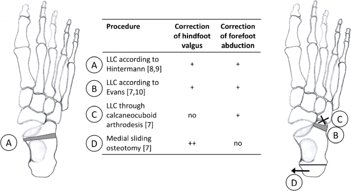
Medial calcaneal slide osteotomy technique. The four most common types are the evans dwyer medial shift and lateral shift. In addition the medial slide calcaneal osteotomy utilizes the same incision as the osteotomy decreasing the need to make a separate incision for screw fixation at the posterior heel. Daniel abstract the medializing calcaneal osteotomy is a frequently performed procedure usually done in conjunction with a flexor tendon transfer as part of a flatfoot correction surgery. 25 medializing calcaneal osteotomy brian s.
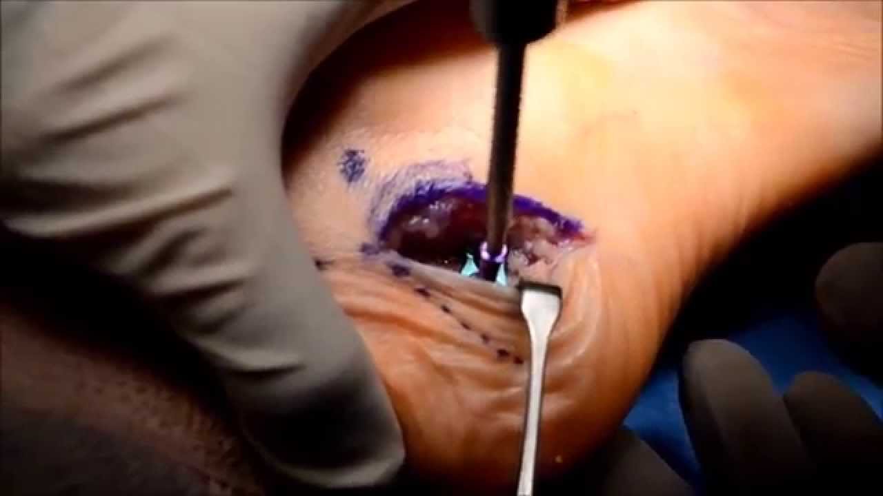
Although further prospective randomized clinical trials are needed early weightbearing may be possible with the use of locking plate fixation for medial slide calcaneal osteotomies. Traditionally the osteotomy is performed using a direct lateral or extended lateral approach which may carry the risk of wound problems infection and neurovascular injury. The incision was carried down through the skin only with a 15 blade knife. A calcaneal osteotomy comprises of making a cut across the heel bone and shifting it toward the inside medial or outside lateral.
This article reviews indications techniques complications and outcomes for the medializing calcaneal osteotomy. In this setting plantar fascia release has traditionally been performed through an incision over the medial calcaneal tuberosity and the calcaneal osteotomy through a lateral incision. In a calcaneal osteotomy an incision is made on the outer or lateral side of the foot. Calcaneal osteotomy cadaveric lab with nicholas abidi md dr.
Jordan grossman demonstrates a medial displacement calcaneal osteotomy using fixos 2. This animation represents an extract of the surgical technique. Winters and joseph n. Dr performed a lateral slding calcaneal osteotomy along with a lateral column lengthening need help with cpt code.
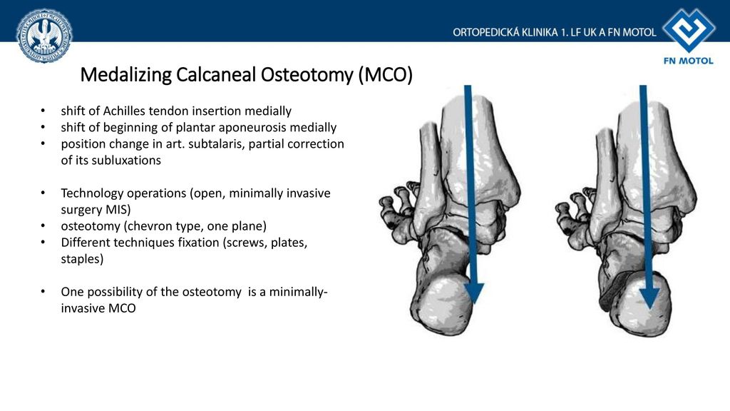
Nicholas abidi performs a medial calcaneal osteotomy to correct an adult flatfoot deformity using large acutrak 2 7 5 screws. The rationale for the use of the screw radiographic evidence and surgical steps are provided. By sliding the posterior tuber of the calcaneus 1 cm medially the mechanical axis of the limb is. Medial displacement calcaneal osteotomy is a common procedure often used as part of pes planovalgus deformity correction.
Plantar fascia release and calcaneal slide osteotomy are often components of the surgical management for cavovarus deformities of the foot. The heel bone called the calcaneus is the main bone that lies in the heel of the hindfoot. Medializing calcaneal osteotomy is the workhorse operation for correction of hindfoot valgus reliably correcting deformity with a relatively low complication risk.

Medial Calcaneal Sliding Osteotomy

Medial Calcaneal Sliding Osteotomy Surgery In Washington D C Maryland And Northern Virginia
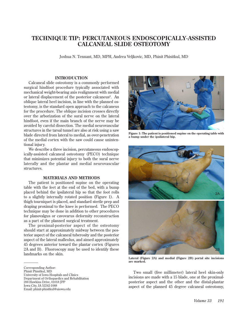
Pdf Technique Tip Percutaneous Endoscopically Assisted Calcaneal Slide Osteotomy

Medial Calcaneal Sliding Osteotomy Lewiston Orthopedics Lewiston Id
Http Citeseerx Ist Psu Edu Viewdoc Download Doi 10 1 1 1026 8013 Rep Rep1 Type Pdf
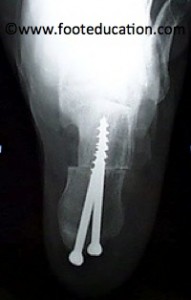
Calcaneal Osteotomy Footeducation

Wheeless Textbook Of Orthopaedics

Medializing Calcaneal Osteotomy Youtube

Arthrex Calcaneus Step Plate

Wheeless Textbook Of Orthopaedics

Paragon28 Calc Slide Plate Orthopedic Implants Paragon28

Key Insights On The Percutaneous Calcaneal Displacement Osteotomy Podiatry Today

Medial Sliding Osteotomy Of The Calcaneus A 59 Year Old Male With Download Scientific Diagram
Http Www Apma Org Files Mdco 20apma Pdf
Flat Foot Anatomy And Current Treatment Ppt Download

Calcaneal Sliding Osteotomy Stryker
Medial Sliding Displacement Calcaneal Osteotomy With Plate Flat Foot Reconstruction Youtube

Figure 1 From Comparison Of Lateral Opening Wedge Calcaneal Osteotomy And Medial Calcaneal Sliding Opening Wedge Cuboid Closing Wedge Cuneiform Osteotomy For Correction Of Planovalgus Foot Deformity In Children Semantic Scholar

Medial Displacement Calcaneal Osteotomy Youtube
Https Kundoc Com Download Effects Of Medial Displacement Calcaneal Osteotomy And Calcaneal Z Osteotomy On 5ac67c10d64ab2e058161e5d Html

Figure 2 From Calcaneus Osteotomy Semantic Scholar

Flatfoot Surgery Musculoskeletal Key

The Photograph A And Standing Lateral Radiograph B Of Left Foot Download Scientific Diagram

Arthrex Calcaneal Osteotomy For Flatfoot
Https Encrypted Tbn0 Gstatic Com Images Q Tbn 3aand9gcrzcauc5ccpdxgi1ophbi6ez Sewcgsaukt6anxsmfg2gb1gjvt Usqp Cau

Figure 5 From What Is The Role And Limit Of Calcaneal Osteotomy In The Cavovarus Foot Semantic Scholar

Current Concepts With The Evans Calcaneal Osteotomy And Lateral Column Lengthening Podiatry Today

Medial Calcaneal Sliding Osteotomy Resurgens Orthopaedics

Figure 1 From Concomitant Calcaneo Cuboid Cuneiform Osteotomies And The Modified Kidner Procedure For Severe Flatfoot Associated With Symptomatic Accessory Navicular In Children And Adolescents Semantic Scholar

Key Pearls Of Calcaneal Osteotomies Podiatry Today
Http Aoj Amegroups Com Article Viewfile 5681 Pdf

Lateral Column Lengthening By Calcaneal Osteotomy Techniques In Foot Ankle Surgery
Functional Limitations After Lateral Column Lengthening Osteotomy Of The Calcaneus Are Associated With Lower Quality Of Life Springerlink
Https Www Ankleandfootcare Com Research Jfas Dco Pdf
Https Journals Sagepub Com Doi Pdf 10 1177 1071100717712543

Gastrocnemius Recession Intramuscular Approach Lewiston Orthopedics Lewiston Id
Http Www Podiatryinstitute Com Pdfs Update 2002 2002 11 Pdf
Https Www Acfas Org Uploadedfiles Physicians Member Center Acfas Regional Divisions Divisions Post2014 Pdf 1177 Sci928 Pdf
Http Www Podiatryinstitute Com Pdfs Update 2003 2003 42 Pdf

Pdf Calcaneal Osteotomy Safe Zone To Prevent Neurological Damage Fact Or Fiction

Normal Appearance Of Medial Displacement Calcaneal Osteotomy A Download Scientific Diagram
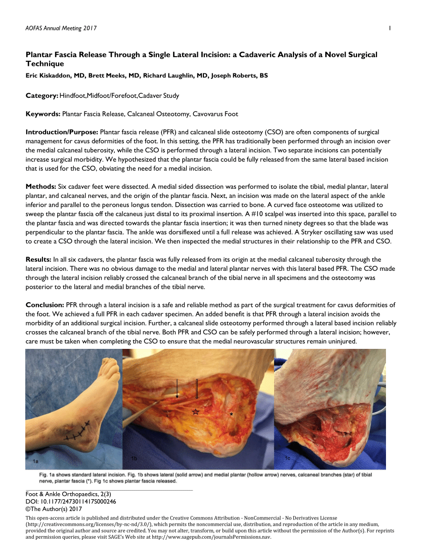
Pdf Plantar Fascia Release Through A Single Lateral Incision A Cadaveric Analysis Of A Novel Surgical Technique
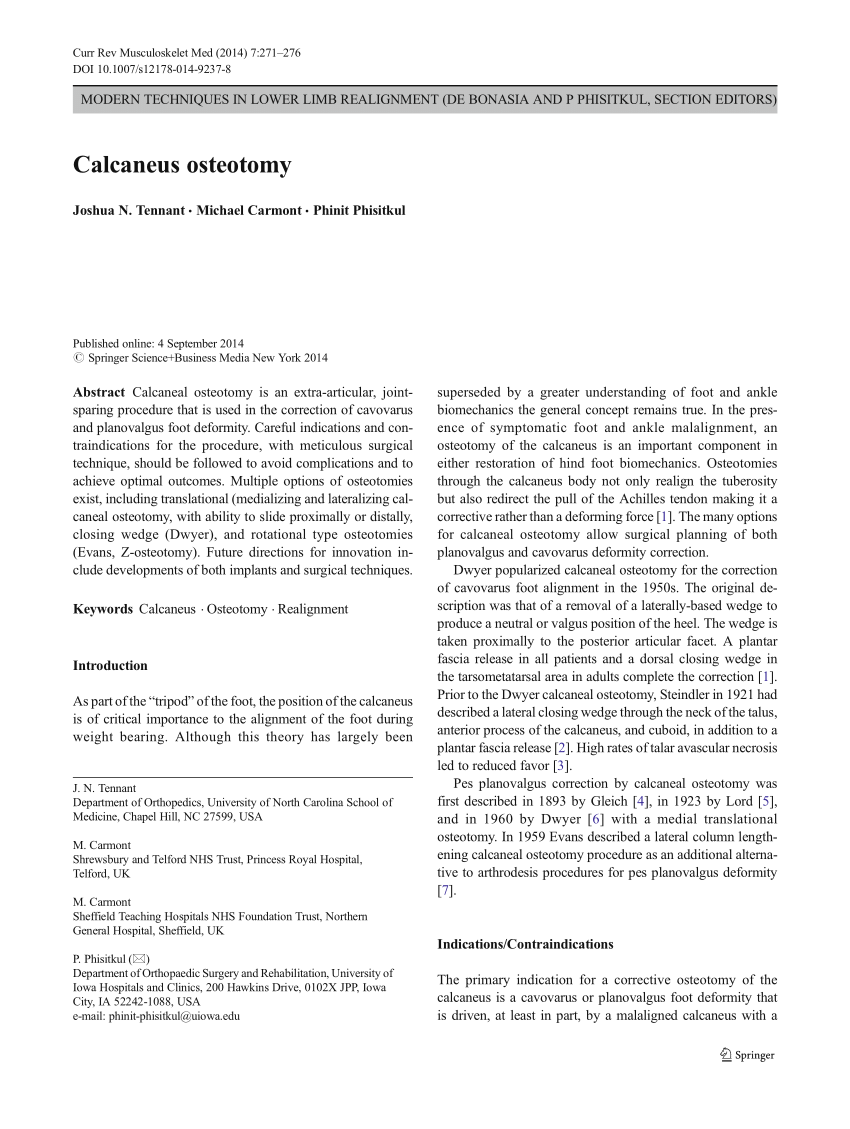
Pdf Calcaneus Osteotomy
Https Www Researchgate Net Profile Scott Mubarak Publication 13465837 Calcaneal Cuboid Cuneiform Osteotomy For The Correction Of Valgus Foot Deformities In Children Links 59f772a2aca272607e2d837d Calcaneal Cuboid Cuneiform Osteotomy For The Correction Of Valgus Foot Deformities In Children Pdf

Pdf Calcaneal Osteotomy And Transfer Of The Tendon Of Flexor Digitorum Longus For Stage Ii Dysfunction Of Tibialis Posterior Three To Five Year Results

Nonunion The Foot And Ankle Online Journal
Http Citeseerx Ist Psu Edu Viewdoc Download Doi 10 1 1 893 1439 Rep Rep1 Type Pdf